|
Robert Lufkin’s Twitter followers approaches 30,000 as he provides key updates to the COVID-19 pandemic situation. Beginning in 2019 he was able to grow his Twitter audience from zero to >20,000 followers in about 180 days. The account at https://twitter.com/robertlufkinmd is described as ‘Med school professor exploring #COVID19, #artificialintelligence, #genetics, #consciousness, tech, optimal #health, #longevity, quantified self, #DIY, & more‘. It presently has >2500 tweets and up to one million views a month in traffic. Robert Lufkin has served as a tenured medical school professor for decades and is author of over 200 peer reviewed scientific papers, 32 chapters and 13 books translated into four languages on medical imaging. He has an international reputation for excellence, has given invited lectures/keynotes around the world and was named one of the 100 most creative people in Los Angeles by Buzz Magazine. His honors include serving as President of the Society of Magnetic Resonance Imaging, President of the American Society of Head and Neck Radiology, and numerous other professional affiliations. Among his many inventions including patents in artificial intelligence, he developed and helped commercialize an MR-compatible biopsy needle which is sold and used worldwide today as the “Lufkin Needle.” He has also founded and/or invested in several technology companies. This diverse experience has paid off when he ventured into his current social media. Robert Lufkin has had a lifelong interest in neuroscience and artificial intelligence. During high school in the Boston area, he was fortunate to be able to attend classes at the Massachusetts Institute of Technology [MIT]. During that time he also worked at the New England Primate Center affiliated with Harvard University where he helped in the laboratories of David Hubel and Torsten Wiesel who studied the mammalian visual system. They later received the Nobel Prize for their work describing the neurophysiology of the visual cortex. In college at Brown University, Robert did original experimental research in the neurophysiology of the mammalian visual system, specifically the superior colliculus. He also minored in computer science and worked part-time in the main campus computer center as a machine operator to earn extra money. Robert next attended the University of Virginia School of Medicine, where he also studied computer science in healthcare at the National Institutes of Health in Bethesda, Maryland. He completed an internship in internal medicine at the University of Oregon Center for Health Sciences. He completed a residency in diagnostic radiology [where he served as Chief Resident] at the University of California, Los Angeles [UCLA] School of Medicine. After residency he did a fellowship in Neuroradiology/Head and Neck Radiology with Dr. William Hanafee and was recruited to join the faculty at UCLA where he eventually became a tenured Professor of Radiology. 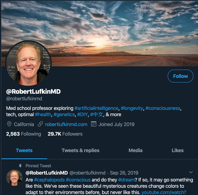
7 Comments
RAVEN MRI TEACHING FILE) Edited by Robert Lufkin William Bradley and Michael Brant-Zawadski includes Dr Lufkin’s book on MRI of the Head and Neck which was based on Dr Lufkin’s popular UCLA Visiting Fellowship. Visitors come from all over the world to learn the basics of MRI as well as its practical clinical applications in the head and neck and other areas from Dr Lufkin and his team. Dr Lufkin is internationally recognized for his expertise in MRI and Head and Neck. He has hundreds of publications on basics of MRI as well as its clinical applications in the head, neck and brain. He also was elected and served as President of the international Society of Magnetic Resonance Imaging as well as the American Society of Head and Neck Imaging. In addition to running the MRI visiting fellowship, continuing with cutting edge MRI clinical research, and seeing patients, Dr Lufkin also speaks all over the world on various topics related to MRI. Dr Lufkin also served as Chief of Head and Neck Radiology at UCLA School of Medicine for two decades. The First Edition of this text is part of the popular Raven MRI Teaching File Series. The book presents 100 actual case studies that cover a wide range of head and neck disorders and demonstrate the use of current MRI techniques and contrast enhancement agents to aid in diagnosis. Each case study is illustrated with high-resolution MR images and presented in an easy-to-follow format on a two-page spread. On the left-hand page are the images and the clinical history. On the right-hand page are concise descriptions of the radiographic findings, the diagnosis, and the pathology. This format is ideal for teaching readers how to interpret MR images or for everyday reference at the view box. Praise this edition:"The book is packed with pearls....It should be useful to residents and generalists in radiology and to those in other disciplines in which MR imaging is used....This book is well worth the investment."--American Journal of Roentgenology The entire series now available on CD-ROM contains 10 volumes of material related to MRI. 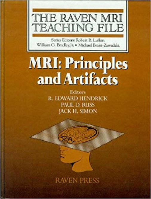 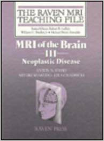 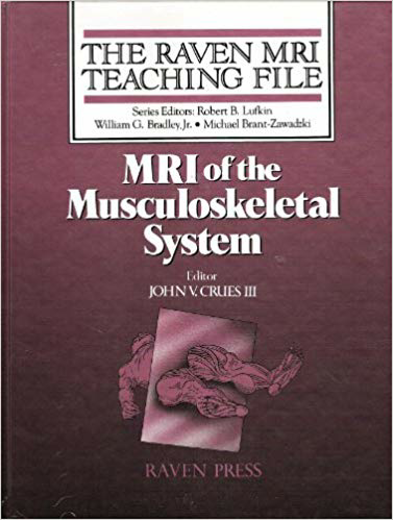     Open Field MRI edited by Robert B Lufkin and DHW Gronemeyer is based on Dr Lufkin’s popular UCLA Visiting MRI Fellowship. Visitors come from all over the world to learn the basics of MRI as well as its practical clinical applications in the head and neck and other areas from Dr Lufkin and his team. Dr Lufkin is internationally recognized for his expertise in MRI and Head and Neck. He has hundreds of publications on basics of MRI as well as its clinical applications in the head, neck and brain. He also was elected and served as President of the international Society of Magnetic Resonance Imaging as well as the American Society of Head and Neck Imaging. In addition to running the MRI visiting fellowship, continuing with cutting edge MRI clinical research, and seeing patients, Dr Lufkin also speaks all over the world on various topics related to MRI. Dr Lufkin also served as Chief of Head and Neck Radiology at UCLA School of Medicine for two decades. Dr Lufkin’s book covers issues related to the practice of both diagnostic and interventional MRI on this new type of open design MR scanner. The unique configuration and often mid-field strengths of these machines necessitates new strategies for both diagnostic and interventional procedures compared to that of the standard 1.5 tesla diagnostic-only MR scanners, to which the majority of the radiologic literature is addressed today. This broad, multi-authored work will appeal to radiologists, medical physicists, other physicians, and health care personnel. ISBN-13: 978-3540637813 ISBN-10: 3540637818 https://www.amazon.com/Open-Field-Magnetic-Resonance-Imaging/dp/3540637818/ref=sr_1_10?keywords=interventional+MRI&qid=1575067951&s=books&sr=1-10   |